Showing posts with label Science. Show all posts

BioMolecules : Applications of Recombinant DNA

Human Cells with a level of assessment for Laws. Just give the analysis of DNA, RNA and others.

Central Dogma of Molecular Biology
DNA (gene)  ->  RNA (transcript)  ->  Protein (trait)

Different organisms have different traits based on their genes (DNA sequences). For example, frogs have antimicrobial peptides on their skin. Some jellyfish have proteins that allow them to glow in the dark. Mutations in hemoglobin genes lead to anemia.

Based on the central dogma, if transcription and translation of genes lead to some traits, then the insertion of certain genes in a given organism may provide it with new traits. This is the basis for the development of genetically modified organisms (GMOs).

Presentation of Recombinant DNA

There are many different traits that can be introduced to organisms to change their properties. The following table shows examples of modified traits using cloned genes and their applications:


PCR Amplification

Once a desired trait is chosen, information must be acquired for either its detection or expression in a given organism.

Detection
Some researchers may be interested in determining if a given gene/trait is available in a particular organism. If no previous research provides this information, researchers may test the DNA of different organisms for the presence of these specific genes. A technique that allows the detection of specific genes in target organisms is called PCR.

PCR amplification is an in-vitro method that simulates DNA replication in vivo. It utilizes a thermostable (heat-resistant) DNA polymerase that builds single stranded DNA strands unto unwound DNA templates. PCR uses repeated cycles of incubation at different temperatures to promote the unwinding of the DNA template (~95°C); the annealing of a primer (a ~20bp oligonucleotide sequence (recall RNA primers in DNA replication) onto the ssDNA template strand (~54 - 60°C); and the extension of the generated ssDNA strand through the binding of complementary bases to the template strand (~72° C).

The thermostability of the polymerase allows it to survive the repeated cycles of denaturation, annealing and extension with little loss of enzyme function. Each cycle of PCR doubles the amount of the target sequence. A typical PCR experiment uses about 35 cycles of amplification. This increases the original amount of the target sequence by 235 (i.e. ~34 billion) times. Gene detection by PCR involves the design of primers that would only bind to sequences that are specific to a target. For example, researchers would want to find out if gene X (e.g. the gene for insulin) is available in a target organism (e.g. a mouse, Mus musculus). Primers may be designed by looking at the available sequences for gene X in the databases (e.g. all the genes for insulin in different organisms; humans, pigs, cows, etc.).

The different gene X sequences must be aligned/ compared to match areas of sequence similarity (conserved sequences) and areas of sequence dissimilarity (non-conserved sequences). Primers designed to have the same sequence as the conserved areas will be specific for binding gene X sequences in all the target organisms. Primers designed to have the same sequence as the non-conserved areas will only be specific for the organisms which match its sequence.

Primers may be classified as forward or reverse primers. Forward primers are complementary and bind to the reverse complementary (non-coding) sequence of the gene. Reverse primers are complementary and bind to the coding sequence of the gene.

PCR Applications

1. PCR may be used to detect the presence of a desired gene in an organism. Depending on the primer design, the expected product may represent only a specific region of the gene or the entire gene itself. The first case is useful for detection of the gene, or the detection of organisms with that specific gene within a sample. The second case is useful for the amplification of the entire gene for eventual expression in other organisms. The direct amplification/copying of a full gene is part of the process for “cloning” that gene.

2. Cloning and Expression
Some genes provide economically, and industrially important products (e.g. insulin-coding genes; genes for collagen degradation). In some cases, scientists would want to put these genes into organisms for the expression of their products. One example would be the insertion of an insulincoding gene from the human genome into bacteria. This allows the “transformed” bacteria to now produce human insulin as a product.

Certain types of bacteria are capable of this process since they are able to take genes within their cell membranes for eventual expression. The genes are normally in the form of small, circular DNA structures called plasmids.

The genes found in the inserted plasmid DNA sequence will be expressed as proteins that provide specific traits to the transformed bacteria. The basic components of an expression plasmid are listed in the following table. The purpose of each of these is also provided.

There are certain ethical principles should be followed and adhered to in the production of genetically modified organisms. Animal welfare should be taken cared of and human cloning must never be conducted.

Lesson 1 - BioMolecules : Structure and Functions
Lesson 2 - BioMolecules : DNA Replication and Protein Synthesis
Lesson 3 - BioMolecules : Genetic Engineering 

Read More......

BioMolecules : Genetic Engineering

Human Cells with a level of assessment for Laws. Just give the analysis of DNA, RNA and others.

Genetic Engineering
1. Classical breeding practices focus on the mating of organisms with desirable qualities.
2. Genetic engineering involves the use of molecular techniques to modify the traits of a target organism. The modification of traits may involve:

      I. introduction of new traits into an organism
      II. enhancement of a present trait by increasing the expression of the desired gene
      III. enhancement of a present trait by disrupting the inhibition of the desired genes’ expression.
3. A general outline of recombinant DNA may be given as follows:

      I. cutting or cleavage of DNA by restriction enzymes (REs)
      II. selection of an appropriate vector or vehicle which would propagate the recombinant DNA (  eg. circular plasmid in bacteria with a foreign gene of interest)
      III. ligation (join together) of the gene of interest (eg. from animal) with the vector ( cut bacterial plasmid)
      IV. transfer of the recombinant plasmid into a host cell (that would carry out replication to make huge copies of the recombined plasmid)
      V. selection process to screen which cells actually contain the gene of interest
      VI. sequencing of the gene to find out the primary structure of the protein

4. Ways in which these plasmids may be introduced into host organisms.

Biolistics. In this technique, a “gene gun” is used to fire DNA-coated pellets on plant tissues. Cells that survive the bombardment, and are able to take up the expression plasmid coated pellets and acquire the ability to express the designed protein.

Plasmid insertion by Heat Shock Treatment. Heat Shock Treatment is a process used to transfer plasmid DNA into bacteria. The target cells are pre-treated before the procedure to increase the pore sizes of their plasma membranes. This pretreatment (usually with CaCl2) is said to make the cells “competent” for accepting the plasmid DNA. After the cells are made competent, they are incubated with the desired plasmid at about 4°C for about 30min. The plasmids concentrate near the cells during this time. Afterwards, a “Heat Shock” is done on the plasmid-cell solution by incubating it at 42°C for 1 minute then back to 4°C for 2 minutes.

The rapid rise and drop of temperature is believed to increase and decrease the pore sizes in the membrane. The plasmid DNA near the membrane surface are taken into the cells by this process. The cells that took up the plasmids acquire new traits and are said to be “transformed”.

Electroporation. This technique follows a similar methodology as Heat Shock Treatment, but, the expansion of the membrane pores is done through an electric “shock”. This method is commonly used for insertion of genes into mammalian cells.

5. Some methods to screen recombinant cells are as follows:

Selection of plasmid DNA containing cells
A selection marker within the inserted plasmid DNA sequence allows the selection of “transformants”. Usually, an antibiotic resistance gene (e.g. AMP ampicillin resistance gene) is included in the plasmid DNA. This allows only “transformed” cells to survive in the presence of the antibiotic (e.g. ampicillin). Plating the plasmid-cell solution on antibiotic-containing media will select for these “transformants” and only allow plasmid-containing cells to grow and propagate into colonies.

Selection of transformed cells with the desired gene
Certain inserted genes within the plasmids provide visible proof of their presence. These include the antibiotic resistance genes that allow for the selection of the transformed cells within the solution. Some inserted genes also produce colored (e.g. chromogenic proteins) or fluorescent products (e.g. GFP) that label the colonies/cells with the inserted gene.

In some cases, the location of the cloning site within the plasmid is in the middle of a gene (i.e. β galactosidase, lacZ) that generates a (blue) colored product in the presence of a substrate (i.e. isopropyl β-D-1 thiogalactopyranoside, or IPTG). Cells transformed with these “empty” plasmids will turn blue in the presence of IPTG. Insertion of a gene in the cloning site disrupts the sequence of the β-galactosidase gene and prevents the generation of the colored product in the presence of the substrate. Cells transformed with the disrupted β galactosidase gene will remain “white” in the presence of IPTG. This “blue-white screening” protocol is thus able to screen for cells that were transformed with the desired gene in the cloning site.

PCR detection of plasmid DNA
Alternatively, the presence of the desired gene in the inserted plasmids may be confirmed using PCR amplification. PCR reactions specific for the desired gene may be done using DNA from cells. Amplification of the expected product would confirm the presence of the gene within the samples. PCR reactions specific for plasmid sequences will also confirm/identify the type of plasmid used for the transformation.

Genetically Modified Organisms (GMOs) With the ability to insert gene sequences, comes the possibility of providing new traits for these target organisms. This has allowed the development of GMOs. Some of these genetic modifications promise higher product yield for their targets. These include the Flavr-Savr Tomato and Bt-Corn.

The Flavr-Savr (“Flavor Savor”) tomato was the first genetically modified organism that was licensed for human consumption. The trait modified in this tomato is its ripening process. A gene for an enzyme that causes the degradation of pectin in the cell walls (i.e. polygalacturonase) normally softens the fruit as it ripens. In Flavr Savr tomatoes, an inhibitor (i.e. antisense RNA) disrupts the expression of this gene, thereby delaying the softening of the fruit and extending the time it may be kept in storage and transported to markets.

Bt-Corn was developed to incorporate the production of a toxin (i.e. Bt-endotoxin) from Bacillus thuringensis in corn plants. This toxin results in the death of pests that feed on these plants like the corn borer larvae. The toxin has been shown to be selective for Lepidoptera larvae and is non-toxic to humans, mammals, fish and birds. The selective toxicity of the toxin allows its use in foodcrops. The introduction of the toxin is believed to increase crop production due to decreased losses from pest infestation. The same technology has been applied in the Philippines for the development of Bt-Eggplant.

Despite the proposed benefits of GMOs, some people have raised their concerns regarding the consumption of these modified foods. While most of the products are tested for safety, concerns are raised for the possibility of not being able to detect hazards that are present, but are currently undetectable by today’s current technology.

Because of these issues, manufacturers are urged to provide labels that notify consumers of GMO presence in their products. While GMOs are believed to be safe when licensed by the food regulatory agencies, it is believed that the consumers must be provided with enough information to make their own choices regarding their use.

Lesson 1 - BioMolecules : Structure and Functions
Lesson 2 - BioMolecules : DNA Replication and Protein Synthesis
Lesson 4 - Biomolecules : Applications of Recombinant DNA

Read More......

BioMolecules : DNA Replication and Protein Synthesis

Human Cells with a level of assessment for Laws. Just give the analysis of DNA, RNA and others.
1. DNA replication or DNA synthesis.
 DNA strands separate and serve as templates for the production of new DNA molecules.

     A. The following are features of replication:
          i. Semiconservative- the resulting DNA consists of one old and one new strand
          ii. Base pairing is maintained; Adenine pairs with Thymine, Guanine pairs with Cytosine
          iii. New DNA molecules are produced in the 5’ to 3’ direction
          iv. Semi discontinuous. The leading strand is synthesized in a continuous manner (5’ to 3’) while the lagging strand is produced discontinuously in short stretches called Okazaki fragments.

     B. In lagging strand synthesis, there is a need for a primer terminus which is provided by an RNA molecule. RNA is synthesized by a primase or RNA polymerase. The 3’OH of the RNA is where new DNA nucleotides are added thus new DNA is built in the 5’ to 3’ direction.

     C. Enzymes in replication are as follows:
         1. helicase;
         2. gyrase;
         3. SSB (single strand binding proteins);
         4. primase or RNA polymerase;
         5. DNA polymerase and
         6. DNA ligase.


2. Transcription or RNA synthesis.

DNA is unwound and one strand is used as template for the production of an RNA molecule.
An RNA polymerase makes RNA in the 5’ to 3’ direction.

Specific regions in the DNA called promoters allow the binding of transcription factors which make possible the binding of RNA polymerase.

Three major types of RNA are: messenger RNA (mRNA); transfer RNA (tRNA) and ribosomal RNA (rRNA).


3. Translation or protein synthesis.

This occurs in the ribosome. Basic ingredients are the various types of RNAs produced in transcription and some proteins or enzymes. The mRNA contains triplets of bases called codons that specify an amino acid, eg. UUU-phe. Various tRNAs carry amino acids from the cytoplasm to the actual site of translation in the ribosome. A tRNA has an anticodon that pair with a codon in the mRNA. Different rRNAs combine with ribosomal proteins to make up the subunits of a ribosome. A functional ribosome has a small and a large subunit.

In bacteria, transcription and translation may be simultaneous. In eukaryotic cells, mRNA, tRNA and rRNA travel from the nucleus to the cytoplasm through the nuclear pores. RNAs may undergo processing. Some unnecessary parts like introns are removed. In eukaryotic mRNA, a 5’ cap and a 3’ poly A tail are added. Coding regions of mRNA are called exons. They specify functional protein products.

The genetic code is the correspondence of the mRNA codons to amino acids.

An amino acid is specified by a codon with three code letters. The genetic code is shown as follows:


Here is a 3D animation that shows how proteins are made in the cell from the information in the DNA code.


Practice Exercises :

REPLICATION :

A. Given the following coding sequence for DNA, provide the sequence of the complementary (template) sequence.

Coding sequence : 5’GATTAGGAATGTATGACTACAGATACGTA 3’

( Complete the Answer)
Complementary sequence 3’ CTAA_______________________________________5’

TRANSCRIPTION :

B. Convert the given coding sequence into an mRNA transcript:

Complementary Non-coding/ Template sequence 3’ CAGTATACGATCGTAGACTATGCTGC 5’

(Complete the Answer) 
Coding sequence ~ mRNA transcript 5’ GUC_______________________________ 3’

TRANSLATION :

C. Translate the given mRNA transcript into a polypeptide sequence:

Coding sequence ~ mRNA transcript 5’ AUGCAUAGAUUAGGAUAUCCCAGAUAG 3’

(Complete the Answer)
Polypeptide sequence N - Met - His -___- Leu -___- Tyr -___- Arg - C

Lesson 1 - BioMolecules : Structure and Functions
Lesson 3 - BioMolecules : Genetic Engineering 
Lesson 4 - Biomolecules : Applications of Recombinant DNA

Read More......

BioMolecules : Structure and Function

Human Cells. Provides high level of assessment and understanding with review guides.
DNA - it is the repository of genetic information
RNA - it transcripts. it is the link between the gene and the gene product (protein)
Protein - are functional products. They are executors of cellular functions

The building blocks of any nucleic acid are the nucleotides.

A nucleotide is composed of a phosphate group (with negative charges), a sugar portion and an N-base.

The sugar in DNA is deoxyribose while the sugar in RNA is ribose.

DNA and RNA are polynucleotides. N-bases are either purines or pyrimidines.
Purine bases are Adenine (A) and Guanine (G).
Pyrimidines are Cytosine (C), Thymine (T, in DNA only) and Uracil (U, found only in RNA)

Specific base pairings occur in DNA. A pairs with T; G pairs with C

DNA is double stranded while RNA is single stranded with Uracil instead of Thymine.


Main Functions:

DNA: repository of genetic information; sequence of bases encodes the blueprint for life processes

RNA: information in the form of base sequence is transformed (transcribed) into mRNA, tRNA and rRNA. DNA is the template copied into RNA by base pairing. G with C; A with U.

Protein: functional products of genes; executes cellular functions

The four structural levels of proteins are:

1.Primary- sequence of amino acids in the polypeptide chain;
2. Secondary- when the polypeptide chains form a helix or a pleated sheet structure;
3. Tertiary- coiling of the polypeptide, combining helices and sheet forms;
4. Quaternary- the association of two or more polypeptides in space

Exercises :

REPLICATION :

A. Given the following coding sequence for DNA, provide the sequence of the complementary (template) sequence.

Coding sequence : 5’ ATGCATAGATTAGGATATCCCAGATAG 3’

( Complete the Answer)
Complementary sequence 3’ TACGTATCTAATC______________________5’

TRANSCRIPTION :

B. Convert the given coding sequence into an mRNA transcript:

Complementary Non-coding/ Template sequence 3’ TACGATCTAATCCTATAGGGTCTATC 5’

(Complete the Answer) 
Coding sequence ~ mRNA transcript 5’ AUGCUAGAUUAG_____________________ 3’


Lesson 2 - BioMolecules : DNA Replication and Protein Synthesis
Lesson 3 - BioMolecules : Genetic Engineering 
Lesson 4 - Biomolecules : Applications of Recombinant DNA

Read More......

Pedigree Analysis with Examples

Medical Doctors usually use a pedigree analysis chart to show genetic disorders are inherited in a family. They can use this to determine the probability (chance) that someone in a family will inherit a certain condition.

This is called pedigree analysis. All the family members are mapped onto a family tree.

Example :


Practice Exercises :


A. Is this trait dominant or recessive?

B. What are the most probable genotypes of I-3 and I-4?

C. What are the most probable genotypes of II-4 and II-5?

D. What is the probability that II-4 and II-5 will have another normal offspring?

Read More......

Bio Sci Quiz 2

Higher level of assessment and knowledge. Analysis with tehnology data.
Structure and Functions of Nucleic Acids : DNA RNA Proteins, Nucleotides. Summary of Cell Parts and Function. Definition of Diffusion , Osmosis, Mediated Transport Mechanism, Facilitative Diffusion and Active transport. Comparison of Membrane Transport Mechanisms.


Read More......

Radiometric Dating of Rocks - Absolute or Not ?

Today, scientists obtain the absolute dates of rocks using radiometric methods. These radiometric methods use radioactive minerals in rocks as geological clocks. The principle involved here is that the atoms of some chemical elements have different forms, called isotopes. These isotopes break down over time in a process scientists call radioactive decay.

RADIOACTIVE DECAY


CALCULATING RADIOACTIVE DECAY


ASSUMPTIONS MADE BY SCIENTISTS

Assumption 1: Conditions at Time Zero

With regard to the volcanic lavas that erupted, flowed, and cooled to form rocks in the unobserved past, evolutionary geologists simply assume that none of the daughter argon-40 atoms was in the lava rocks.

For the other radioactive “clocks,” it is assumed that by analyzing multiple samples of a rock body, or unit, today it is possible to determine how much of the daughter isotopes (lead, strontium, or neodymium) were present when the rock formed.

Yet lava flows that have occurred in the present have been tested soon after they erupted, and they invariably contained much more argon-40 than expected.

For example, when a sample of the lava in the Mt. St. Helens crater (that had been observed to form and cool in 1986) (Figure 1) was analyzed in 1996, it contained so much argon-40 that it had a calculated “age” of 350,000 years!

Similarly, lava flows on the sides of Mt. Ngauruhoe, New Zealand, known to be less than 50 years old, yielded “ages” of up to 3.5 million years.


So it is logical to conclude that if recent lava flows of known age yield incorrect old potassium-argon ages due to the extra argon-40 that they inherited from the erupting volcanoes, then ancient lava flows of unknown ages could likewise have inherited extra argon-40 and yield excessively old ages.

There are similar problems with the other radioactive “clocks.” For example, consider the dating of Grand Canyon’s basalts (rocks formed by lava cooling at the earth’s surface). We find places on the North Rim where volcanoes erupted after the Canyon was formed, sending lavas cascading over the walls and down into the Canyon.

Obviously, these eruptions took place very recently, after the Canyon’s layers were deposited. These basalts yield ages of up to 1 million years based on the amounts of potassium and argon isotopes in the rocks. But when we date the rocks using the rubidium and strontium isotopes, we get an age of 1.143 billion years. This is the same age that we get for the basalt layers deep below the walls of the eastern Grand Canyon.

How could both lavas—one at the top and one at the bottom of the Canyon—be the same age based on these parent and daughter isotopes? One solution is that both the recent and early lava flows inherited the same rubidium-strontium chemistry—not age—from the same source, deep in the earth’s upper mantle. This source already had both rubidium and strontium.

To make matters even worse for the claimed reliability of these radiometric dating methods, these same basalts that flowed from the top of the Canyon yield a samarium-neodymium age of about 916 million years,5 and a uranium-lead age of about 2.6 billion years!


Assumption 2: No Contamination

The problems with contamination, as with inheritance, are already well-documented in the textbooks on radioactive dating of rocks. Unlike the hourglass, where its two bowls are sealed, the radioactive “clock” in rocks is open to contamination by gain or loss of parent or daughter isotopes because of waters flowing in the ground from rainfall and from the molten rocks beneath volcanoes. Similarly, as molten lava rises through a conduit from deep inside the earth to be erupted through a volcano, pieces of the conduit wallrocks and their isotopes can mix into the lava and contaminate it.

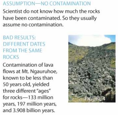
Because of such contamination, the less than 50-year-old lava flows at Mt. Ngauruhoe, New Zealand, yield a rubidium-strontium “age” of 133 million years, a samarium-neodymium “age” of 197 million years, and a uranium-lead “age” of 3.908 billion years!


Assumption 3: Constant Decay Rate

Physicists have carefully measured the radioactive decay rates of parent radioisotopes in laboratories over the last 100 or so years and have found them to be essentially constant (within the measurement error margins). Furthermore, they have not been able to significantly change these decay rates by heat, pressure, or electrical and magnetic fields. So geologists have assumed these radioactive decay rates have been constant for billions of years.

However, this is an enormous extrapolation of seven orders of magnitude back through immense spans of unobserved time without any concrete proof that such an extrapolation is credible. Nevertheless, geologists insist the radioactive decay rates have always been constant, because it makes these radioactive clocks “work”!

New evidence, however, has recently been discovered that can only be explained by the radioactive decay rates not having been constant in the past. For example, the radioactive decay of uranium in tiny crystals in a New Mexico granite yields a uranium-lead “age” of 1.5 billion years. Yet the same uranium decay also produced abundant helium, but only 6,000 years worth of that helium was found to have leaked out of the tiny crystals.


This means that the uranium must have decayed very rapidly over the same 6,000 years that the helium was leaking. The rate of uranium decay must have been at least 250,000 times faster than today’s measured rate! For more details see Don DeYoung’s Thousands . . . Not Billions (Master Books, Green Forest, Arkansas, 2005), pages 65–78.

IF THESE CLOCKS ARE BASED ON FAULTY ASSUMPTIONS AND YIELD UNRELIABLE RESULTS, THEN SCIENTISTS SHOULD NOT TRUST OR PROMOTE THE CLAIMED RADIOACTIVE “AGES.” The assumptions on which the radioactive dating is based are not only unprovable but plagued with problems. As this article has illustrated, rocks may have inherited parent and daughter isotopes from their sources, or they may have been contaminated when they moved through other rocks to their current locations. Or inflowing water may have mixed isotopes into the rocks. In addition, the radioactive decay rates have not been constant.

So if these clocks are based on faulty assumptions and yield unreliable results, then scientists should not trust or promote the claimed radioactive “ages” of countless millions of years.

What do YOU Think ?

Read More......

Vestigial Orgns and Homology

Another branch of comparative anatomy studies structures in humans (and other so called ‘higher’ forms of life) that were believed by evolutionists to be the remains of structures that were required or useful in ‘lower’, less evolved and less complex ancestral forms, but that now no longer are necessary.

In this case, the homologous organ in the more advanced animal is less developed, or even deemed useless. Such homologous structures or organs are referred to as vestigial, with most examples being assumed remnants that resulted from the loss of an earlier, better developed structure. Evolutionists used to proudly point to over a hundred such structures in humans, but the number has decreased consistently as anatomical knowledge has increased.

Today, only a couple of examples at most are usually mentioned (and there is no doubt that even the few examples usually mentioned are useful and not vestigial). As Howitt28 noted, the celebrated German anatomist, Wiedersheim, listed 180 vestigial organs in the human body, but with the increase of knowledge it has been found that every one of them has an important function, although the functions of some organs is presently viewed as minor, or as serving a back-up capacity.

Moreover, if some vestigial organs can be proven to exist, they provide support not for evolution, but for de-evolution|--|i.e. evolution-in-reverse. What the evolutionists must demonstrate is that the development of new and useful organs is occurring today. They also must prove that a process exists that can form new structures called nascent organs, instead of trying to document that once-useful organs now are useless. Evidence for the development of new organs, or those in the process of evolving, would be evidence of evolution. As of now, no evidence of any nascent organ exists.

Evolutionary Naturalism or an Intelligent Designer?

Read More......

Embryology and Homology

One major problem is that in many cases organs and structures which appear identical (or very similar) in different animals do not develop from the same structure or group of embryo cells.

It is not uncommon to find fundamental structures (e.g. the alimentary canal) that form from different embryological tissues in different animals. For example,

in sharks the alimentary canal is formed from the roof of the embryonic gut cavity; in frogs it is formed from the gut roof and floor; and in birds and reptiles it is formed from the lower layer of the embryonic disc or blastoderm.

Even the classic example of vertebrate forelimbs referred to by Darwin (and cited in hundreds of textbooks as proof for evolution) has now turned out to be flawed as an example of homology.

The reason is that the forelimbs often develop from different body segments in different species in a pattern that cannot be explained by evolution.

The forelimbs in the newt develop from trunk segments 2 through 5;
in the lizard they develop from trunk segments 6 to 9;
in humans they develop from trunk segments 13 through 18.

Denton concluded that this evidence shows the forelimbs usually are not developmentally homologous at all.

As an example, he cited the development of the vertebrate kidney which provides a challenge to the assumption that homologous organs are produced from homologous embryonic tissues.

‘In fish and amphibia the kidney is derived directly from an embryonic organ known as the mesonephros, while in reptiles and mammals the mesonephros degenerates towards the end of embryonic life and plays no role in the formation of the adult kidney, which is formed instead from a discrete spherical mass of mesodermal tissue, the metanephros, which develops quite independently from the mesonephros.’

This research supports ReMine’ biotic message theory, the conclusion that the natural world was specifically designed to look like it did not evolve, but was created. ReMine uses a wide variety of examples to support his thesis which has been very favorably reviewed by the creationist community. ReMine notes that homology has been used as evidence against a designer for decades, but as this review shows, it strongly supports the biotic message theory.

Evolutionary Naturalism or an Intelligent Designer?

Read More......

BioChermical Homology

The homology argument from biochemistry parallels the argument in anatomy. Evolutionists suggest that just as the study of comparative anatomy has found evidence of anatomical homologies, likewise research on

“ … the biochemistry of different organisms has revealed biochemical homologies. In fact, the biochemical similarity of living organisms is one of the most remarkable features of life … .

Cytochrome enzymes are found in almost every living organism: plant, animal and protist. The enzymes of the citric acid cycle are also almost universally distributed.

Chlorophyll a is found in all green plants and almost all photosynthetic protists.

DNA and RNA are found in every living organism and, so far as we can determine, contain the same hereditary coding mechanism.

The fact that underneath the incredible diversity of living things lies a great uniformity of biochemical function is difficult to interpret in any other way but an evolutionary one. Presumably these molecules were put to their current use very early in the history of life and almost all modern forms have inherited the ability to manufacture and use them.”

The fact that animals are ‘so similar in their chemical make-up’ has long been used to support Darwinism.

But extensive biochemical research has revealed that the simplest reason for biochemical homology is that all life requires similar inorganic elements, compounds and biomolecules; consequently, all life is required to use similar metabolic pathways to process these compounds.

Most organisms that use oxygen and rely on the metabolism of carbohydrates, fats and proteins must use a Citric acid Cycle which is remarkably similar in all organisms.

Furthermore, the metabolism of most proteins into energy produces ammonia, which is processed for removal in similar ways in a wide variety of organisms. What evolutionists must explain is why billions of years of evolution have not produced major differences in the biochemistry of life.

Many biochemical structures/systems in yeasts and other so-called ‘primitive life’ forms are almost identical to the biochemical families used in humans. With some minor variations, all life uses the same sugar and lipid family, the same 20 amino acids, about 14 vitamins and the same basic genetic code.

Even the complex proteins used in all life are often identical or very similar. Correspondence even exists between very different forms of life such as prokaryotes and eukaryotes. Ribosomes from bacteria, even though translation signals and other differences exist, have enough similarity that they can be made to ‘translate human messenger RNAs into human proteins—and vice versa’.39 The problem for evolutionists is that the biochemistry of all life, even that allegedly separated by hundreds of millions of years of geologic time and evolution, is too similar. Despite the many significant differences between the two basic cell forms (eukaryotes and prokaryotes), they are both

“ … remarkably similar on the biochemical level … . Procaryotes and eucaryotes are composed of similar chemical constituents. With a few exceptions, the genetic code is the same in both, as is the way in which the genetic information in DNA is expressed. The principles underlying metabolic processes and most of the more important metabolic pathways are identical. Thus, beneath the profound structural and functional differences between procaryotes and eucaryotes, there is an even more fundamental unity: a molecular unity that is basic to life processes.”

Although many biochemical similarities exist in life, millions of biochemical differences exist that are inexplicable via evolution. Many of these differences do not provide a selective advantage as implied by the claim that Darwinistic mechanisms have fine tuned life for the past 3.6 billion years.

Creationists suggest that such differences exist due to the need for ecological balance and because the Creator chose to employ variety. Also, were one compound in an organism to be altered, scores of other compounds with which it interacts would often also need to be changed so that the entire biological system could function as a harmonious unit.

Evolutionary Naturalism or an Intelligent Designer?

Read More......

Genetics and Homology

According to the evolutionary theory, homologous features are programmed by similar genes. Gene sequence similarity would indicate common ancestry since such similarities are unlikely to originate independently through random mutations. If the bones of the human arm evolved from the same precursors as the wing of a bat and the hoof of a horse as evolution teaches, then we should be able to trace these alleged homologies to the DNA that codes for them. Some geneticists thought this knowledge would allow them to find the chemical formula needed to produce an arm, leg, or other structure. But once biologists acquired a greater understanding of genetics, they found that what are labeled as homologous structures in different species often are produced by quite different genes.

Homology predicted that features produced by similar genetic sequences are phylogenetically homologous. There are now so many exceptions to this prediction that the concept of genetic homology cannot now be said to be a rule, but the exception. The classic example is mutations in certain homeotic genes41 which can cause wholesale changes in morphology such as producing two pairs of wings instead of the normal single pair, or replacing a fly’ antenna with a leg (or can even cause eyes to develop on the fly’ leg). Genes that produce results similar to the homeotic genes for flies’ wings have been found in most other animal kinds, including mammals and humans.

In another example, the gene that controls mouse eye colour also happens to control the mouse’ physical size; but the gene that controls the fruit fly’ eye colour controls not the fruit fly’ size, but female sex organ morphology.43 Although mice and flies share a similar gene (called eyeless) which functions to control their eye development, the fly’ multifaceted eye is profoundly different from a mouse’ mammal eye. In both the fly Antennapedia and mouse eyeless, similar homeotic genes control development of structures which are not homologous by either the post-Darwinian phylogenetic or the classical morphological definition.

The finding that similar genes regulate such radically different structures strongly argues against the concept of homology. So many genes used in higher organisms have multiple effects that Ernst Mayr once suggested that genes which control only a single characteristic are rare or nonexistent. The finding that a consistent one-gene/one-characteristic correspondence does not exist has been a major set back to the Darwinian interpretation of homology. Because evolutionary biologists have failed to provide a biological basis for their homology research findings, Roth concluded ‘that the title of de Beer’ 1971 essay|--|Homology, an unsolved problem|--|remains an accurate description … . The relationships between processes at genetic, developmental, gross phenotypic and evolutionary levels remain a black box’.45 Research at the molecular level has failed to demonstrate the expected correspondence between gene product changes and the organismal changes predicted by evolution. Evolution by DNA mutations ‘is largely uncoupled from morphological evolution’.46 An example of this is the large morphological dissimilarity that exists between humans and chimpanzees despite a high similarity in their DNA. In short we now know:

“ … in general the homology of structures such as organs or modules cannot be ascribed to inheritance of homologous genes or sets of genes. Consequently, organ homology cannot be reduced to gene homology. Van Valen recognizes this too and therefore suggests, as an alternative, to reduce homology to a continuity of [developmental] information. Information is not the same as genotypic nucleic acid. But what it is exactly, and how it is continuous, is still an unsolved problem.”

Evolutionary Naturalism or an Intelligent Designer?

Read More......